2023-03-07 00:00:00
KYPHOSCOLIOSIS
Kyphoscoliosis is a condition in which the spine presents an abnormal curvature, visible both in the frontal plane ...


Knee luxation is a less frequent but extremely serious injury, in which the thigh bone (femur) and the calf bone (tibia) lose contact with each other. Most of the times they occur in the case of a car or sports accident or in the case of falling from a height.
Symptoms include knee pain and instability, but also the blood vessels (arteries) in the popliteal area (the area behind the knee) can be injured.
Often, a dislocation of the knee can be accompanied by one or more injuries, such as: fracture, cartilage or torn meniscus, ligaments around the knee torn.
Knee dislocation is often confused with a subluxation, but they are not at all similar, the subluxations being partial dislocations of the knee caused by ligament damage.
The knee is one of the largest and most complex joints of the body, having a complex functionality. The knee joint should be mobile and supple to allow flexion and extension of the lower limb and also stable to support the entire weight of the body.
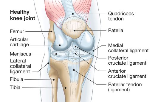
The bone components that form the knee joint are:
➢ the distal extremity of the femur, represented by the two femoral condyles,
➢ the kneecap or the patella, which anteriorly joins the femoral condyles
➢ the proximal extremity of the tibia.
On the edge of the bone surfaces there is articular cartilage, a firm, smooth layer of tissue, lining all the joints of the body. Two cartilage discs in the shape of the letter C, form the meniscuses, laterally and medially, having a role in the absorption of shocks.
The tendons keep the muscles and bones of the knee connected to allow the knee joint to move.
Ligaments join with all the bones of the knee giving it stability. There are 4 ligaments in the knee (2 collateral ligaments located on either side of the knee and 2 cruciate ligaments, anterior and posterior) that act as strong "ropes" to hold the bones together and keep the knee stable.
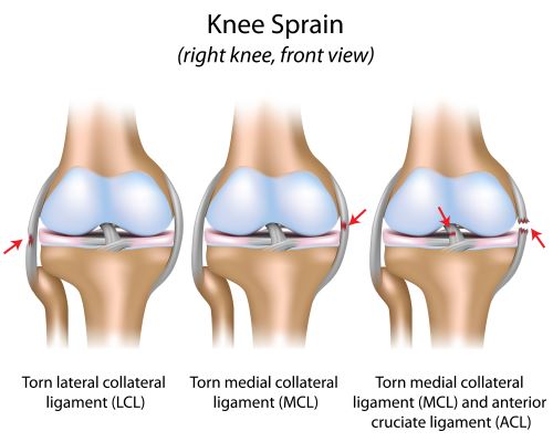
When a dislocation of the knee occurs, there are significant traumas and ligament ruptures, sometimes all the ligaments of the knee can be torn.
Luxations can be:
➢ Anterior, when the femur moves forward on the tibia
➢ Posterior, when the femur moves back on the tibia
➢ Lateral, the femur moves on the lateral side of the tibia
In the early stages of treatment, the priority is the restoration of damaged blood vessels and nerves. In almost all cases, surgery, classical or by arthroscopy, will be required to repair the associated injuries of ligaments, meniscus and cartilage.
Thus, the treatment of knee luxations includes: closed reduction (realignment of bone heads under anesthesia), evaluation of the neurovascular state and surgical restoration of associated injuries (blood vessels, meniscus, ligaments, articular cartilage).
This therapeutic approach, although technically difficult, has significant results in terms of functionality subsequent to the knee joint.
The recovery program through physical therapy is essential for restoring the normal functionality of the knee and for preventing relapses or other injuries.
Initially, light exercises will be performed to mobilize the knee joint, under the guidance of a physical
therapist. Subsequently, the recovery program will continue with exercises to tone the stabilizing muscles of the knee and stretching exercises.
➢ The leg will be raised on a pillow or support, above the level of the heart, in the first days after the injury to help reduce inflammation and swelling of the knee
➢ A knee orthosis will be worn for several weeks after the injury, the orthosis that helps maintain joint stability.
➢ The loading of the sprained knee will be avoided in the first weeks after the injury, walking will be done with the help of crutches.

I always follow professional workflow and provide you the best service with reliable costs.


Kyphoscoliosis is a condition in which the spine presents an abnormal curvature, visible both in the frontal plane ...
The anterior cruciate ligament can suffer injuries when the knee joint is pushed back, twisted or pushed from side ...
Hemiplegia is a condition caused by brain damage or spinal marrow injury that leads to paralysis on one side of the ...