2023-03-06 00:00:00
RETRACTILE CAPSULITIS (FROZEN SHOULDER)
The shoulder joint, also called the scapulo-humeral joint, consists of the humeral head and the scapula (shoulder blade) ...


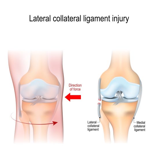
The lateral collateral ligament (LCL), also called the fibular ligament, is the main structure located on the external side of the knee that prevents the knee from slipping to the side and its postero-lateral rotation. It’s a round, thin and strong ligament that is inserted on the side of the femur and on the side of the head of the fibula (peroneum) helping to ensure the stability of the knee.
Lateral collateral ligament injuries can occur in case of sudden stops and/or starts, blows on the internal side of the knee or in case of hyperextension, with or without direct contact. Sometimes, a rupture of the lateral collateral ligament can remain undiagnosed for several weeks, until the instability of the knee begins to be felt. Most commonly, injuries occur on the sports field.
Lateral collateral ligament sprains are less common compared to those of the medial collateral ligament
Initially, the severity of the lesion and the associated complications will be evaluated, in more severe cases an MRI examination or an X-ray will be required.
Healing and protecting ligaments is vital in the case of all injuries, regardless of their severity. In the case of mild and medium ruptures (I and II degree) conservative methods of treatment (without surgery) are preferred, the duration of treatment spanning for up to 8 weeks.
Immediately after an injury, locally applied ice, in sessions of 5-10 minutes, 3-5 times a day, helps to reduce inflammation and pain, as well as swelling (swelling), if any. Always put a thin towel between the ice and the area on which it is applied.
Anti-inflammatory drugs help reduce pain and inflammation, but will be administered for periods of time not exceeding 2 weeks.
Activities that cause pain or that may accentuate the injury will be interrupted. The affected leg will be kept elevated (raised) on a pillow or support, above the level of the heart.
When walking is possible, it will be done with the help of an orthosis that will protect the knee from slipping to the side. The orthosis will be worn for about 6 weeks.
Through specific exercise programs, initially passive and then active, physical therapy significantly helps to restore the injured ligament, aiming at:
➢ prevention of stiffness
➢ toning (strengthening) the stabilizing muscles of the knee
➢ restoring balance
➢ regaining flexibility of the joint through stretching exercises of the quadriceps muscle
➢ stimulation of proprioception which represents the acceleration of the voluntary motor response by stimulating the proprioceptors in the muscles, tendons and joint.
Sports activities can generally be resumed after about 4 weeks in case of mild ruptures (1st degree) and after about 10 weeks in the case of medium ruptures (2nd degree), under the following circumstances:
➢ movement in the knee joint is complete and painless
➢ lateral sensitivity of the knee is reduced
➢ there is no ligament laxity
Reconstruction of the injured ligament may be necessary in case of severe, 3rd degree ruptures. After the intervention, avoid loading (bearing body weight) the affected knee. It is also recommended to wear an orthosis for 4-6 weeks and start the recovery program through physical therapy to prevent joint stiffness and to restore stability and functionality of the knee.
Insist on toning the quadriceps muscle.
Toning the hamstrings muscles should be avoided for a while, for about 4 months, in order to prevent damage to the lateral collateral ligament reconstruction.
Sports activities can be easily resumed after 4 months postoperatively.

I always follow professional workflow and provide you the best service with reliable costs.


The shoulder joint, also called the scapulo-humeral joint, consists of the humeral head and the scapula (shoulder blade) ...
The calf is the part of the foot located between the knees and the foot. The skeleton of the calf consists of two bones ...
The shoulder is composed of several joints, tendons and muscles that come together to allow for a wide ...