2023-03-07 00:00:00
WHY DO CALVES HURT?
The calf is the part of the foot located between the knees and the foot. The skeleton of the calf consists of two bones ...


Peroneal tendinitis is a common cause of pains that occur on the side of the ankle, due to injuries or damage to the peroneal tendons.
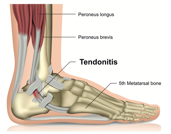
The peroneal tendons are strong fibrous structures that connect the calf muscles with the bones of the foot instep (connects the calf muscles to the bones of the foot). They help stabilize and balance the foot and ankle, protecting them from injuries and/or lesions.
Peroneal tendinitis is the inflammation of the peroneal tendons, as a result of overwork, as it happens in sports that involve intense movements of the ankle (athletics, tennis etc.) or as a result of an acute injury (ankle sprain).
Peroneal tendinitis is not as common compared to other types of leg tendinitis, such as Achilles tendonitis (inflammation of the Achilles tendon).
Other situations that favor the inflammation of the peroneal tendons are:
➢ lack of stretching (stretching) before physical activity
➢ certain conditions, such as diabetes, osteoarthritis, rheumatoid arthritis or gout.
➢ previous lesions of the peroneal tendons
➢ obesity
➢ stiffness of the tendons
➢ arched leg (it is the opposite of the flat foot where the arch of the foot, that is, the portion on the inside of the foot between the heels and the base of the big toe, is taller than normal).
The most common peroneal tendonitis is manifested with ankle pain, especially felt along the length of the affected tendon, pain that worsen during the practice of physical exercise.
Other symptoms include redness, heat and edema (swelling) around the tendon.
Sometimes the tendon may feel thickened.
Untreated, peroneal tendonitis can progress to a partial or complete tendon rupture. Damaged or weakened tendons can also lead to their subluxation. Subluxation of a tendon is a disorder that involves an elongation, rupture or avulsion (plucking) of the tendon.
Torn or subluxed tendons cause: weakness in the ankle or even instability and intense pain along the outer part of the foot and ankle.
Peroneal tendinitis can be difficult to diagnose because the symptoms are similar to those of other foot and ankle problems, such as sprains, arthritis and fractures. Sometimes imaging (X-ray, musculoskeletal ultrasound or MRI examination) may be necessary to confirm if the tendon is the one affected or there are other conditions such as: leg fracture, osteoarthritis, cartilage damage or damaged tissue.
To protect the affected ankle, a special boot or orthosis can be worn that will stabilize the ankle and protect it while walking.
The activities and movements that cause or accentuate the pain will also be avoided. During rest, the affected leg will rise on a support or pillow, preferably above the level of the heart. Wearing a compression bandage around the ankle will reduce its swelling.
Nonsteroidal anti-inflammatory drugs (ibuprofen, diclofenac etc.) in the form of tablets or local ointments help relieve pain and inflammation.
In cases where the pain is not improved, steroid injections (stronger anti-inflammatories) can be administered around the tendon or in the tendon sheath.
Locally applied ice, in sessions of 15-20 minutes, 3-5 times/day, helps significantly reduce inflammation. Always put a thin towel between the ice and the area on which the ice is applied or wrapped in a thin towel, so as not to irritate the skin.
Under the guidance of a physical therapist, stretching exercises will be practiced to restore flexibility in the ankle and foot, as well as strength exercises to increase the stability and strength of the affected foot.
After about a month, all daily activities will be gradually resumed.
If peroneal tendonitis does not improve with conservative treatments (those described above), surgery may be required, which consists in cleaning the damaged outer layers of the tissue from the peroneal tendons.
After surgery, a protective orthosis of the lower leg is worn for 4-6 weeks, the orthosis can be removed for the hygiene of the area and in the first weeks walking can be done with the help of crutches to protect the affected ankle.
Physical therapy after surgery is necessary to regain the strength and stability of the ankle.
➢ in general, in all activities that involve movement, the intensity of physical effort is gradually increased
➢ body weight is maintained within normal limits in order not to put additional pressure on the knees and the lower leg
➢ physical effort is interrupted when pain occurs in the ankle or foot
➢ rest between workouts is essential
➢ insist on warm-up and stretching before starting any physical activity
➢ with the recommendation of a specialist support devices and protection of the ankle and foot will be worn (for example, personalized shoes pads if the arch of the foot is high or supporting orthotics for the ankle in case of an unstable ankle).

I always follow professional workflow and provide you the best service with reliable costs.


The calf is the part of the foot located between the knees and the foot. The skeleton of the calf consists of two bones ...
Spina bifida is a birth defect, which occurs when the spine and spinal marrow are not formed properly ...
The cervical spine is composed of seven vertebrae that stretch from the base of the skull to the upper part of the ...