2023-03-29 00:00:00
PROLAPSE OF THE PELVIC ORGANS
Prolapse is a condition in which the organs fall or slide from their place, most exposed being the pelvic organs ...


The articular cartilage under the kneecap (patella) acts as a natural shock absorber for the knees. When it is injured or overused, it can lead to patellar chondromalacia.
Patellar chondromalacia is a general term that indicates damage to the articular cartilage located under the patella. The main symptom is pain felt in front of the knee, especially during activities that involve squatting, kneeling, climbing stairs, climbing, or simply sitting on a chair.
➢ the natural aging process can facilitate the degeneration of the articular cartilage and implicitly the patellar chondromalacia
➢ wear or the overstrain of the patella also facilitates the degeneration of cartilage
➢ any part of the quadriceps muscle that becomes hypotonic (with reduced muscle tone), can cause a muscular imbalance that also affects the patella
➢ the poorly toned muscles around the hip can indirectly affect the patella and cause joint pain
The knee is one of the largest and most complex joints of the body, having a complex functionality. The knee joint must be mobile and supple to allow flexion and extension of the lower limb and also stable to support the entire body weight.
Walking, running and accelerating suddenly, jumping, twisting on the heel, braking, stopping (more or less sudden) are roles of the knee, which generate numerous pressures, managed by the bone, ligamentous, meniscal and muscular structures of the knee.
The bone components that form the knee joint are:
➢ the distal extremity of the femur represented by the two femoral condyles
➢ the kneecap or patella, which anteriorly unites the femoral condyles
➢ the proximal extremity of the tibia.
On the edge of the bone surfaces there is articular cartilage (a firm, smooth layer of tissue lining all the joints of the body).
Two cartilage discs in the shape of the letter C, form the meniscuses (lateral and medial) that allow the knee to move, but also have a role in the absorption of shocks.
The tendons keep the muscles and bones of the knee connected to allow the knee joint to move.
Ligaments join with all the bones of the knee giving stability to the knee .
When the movement of the knee becomes repetitively abnormal due to a muscle imbalance, the patella may rub irregularly on the femur. The lower surface of the patella becomes inflamed and wears out.
The patella (patella) is usually pulled to the side (outwards) due to a muscular imbalance, namely: either the muscle on the side of the thigh (the vast latera) is stronger, or the muscle on the inside of the scythe (the medial vast) is weaker.

Direct examination and evaluation result in high diagnostic accuracy. An X-ray of the knee may also suggest abnormal wear of the cartilage.
1. Rest
It is essential for restoring the overworked knee joint
2. Anti-inflammatory treatment
It helps reduce pain and inflammation, but is recommended for short periods of time, 7-10 days.
3. Physical therapy
Changing daily activities (giving up repetitive activities that injure the joint) toning the stabilizing muscles of the knee and increasing joint flexibility, are the main objectives of the recovery program.
In the early stages of the disease, exercise programs aim to reduce pain.
Then, as the chondromalacia progresses, the exercise programs pursue:
➢ the toning (strengthening) the muscles located on the anterior side of the thigh (quadriceps)
➢ the stretching of the muscles located on the posterior part of the thigh (femoral biceps, semimembranosus and semitendinosus)
Thus, the forces that keep the kneecap (patella) in a normal, central position will be balanced.
A strong quadriceps muscle also cushions the forces that would otherwise concentrate in the patella.
4. Wearing an orthosis for the patella
Physical exercise help by significantly alleviating pain. On the side, the orthosis is provided with small pads that prevent the patella from slipping to the side.

I always follow professional workflow and provide you the best service with reliable costs.


Prolapse is a condition in which the organs fall or slide from their place, most exposed being the pelvic organs ...
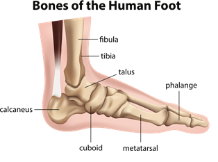
Metatarsalgia is the pain at the plantar level (sole of the foot) under the fingers, at the level of the metatarsal bones ...
Pelvic pain is localized in the lower abdomen and pelvis and can manifest itself as a symptom of diseases of the ...